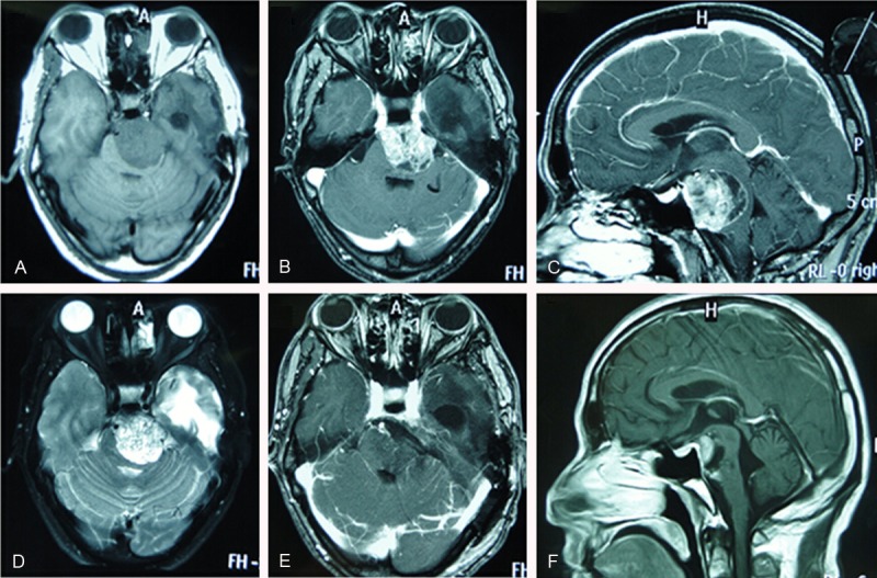
Figure 1.

Radiologic characterization of extra-axial petroclival ependymoma. A, D. Preoperative cranial MRI showing the tumor is hypointensesity on T1-weighted imaging (T1W) and iso- to hyperintensity on T2-weighted imaging (T2W). B, C. Axial and sagittal contrast enhanced T1-weighted MR imaging showing heterogenous enhancement. E. Postoperative axial contrast enhanced T1-weighted MR image revealing the tumor was totally resection. F. Postoperative cranial MRI at 4-month follow-up showing tumor recurrence.
