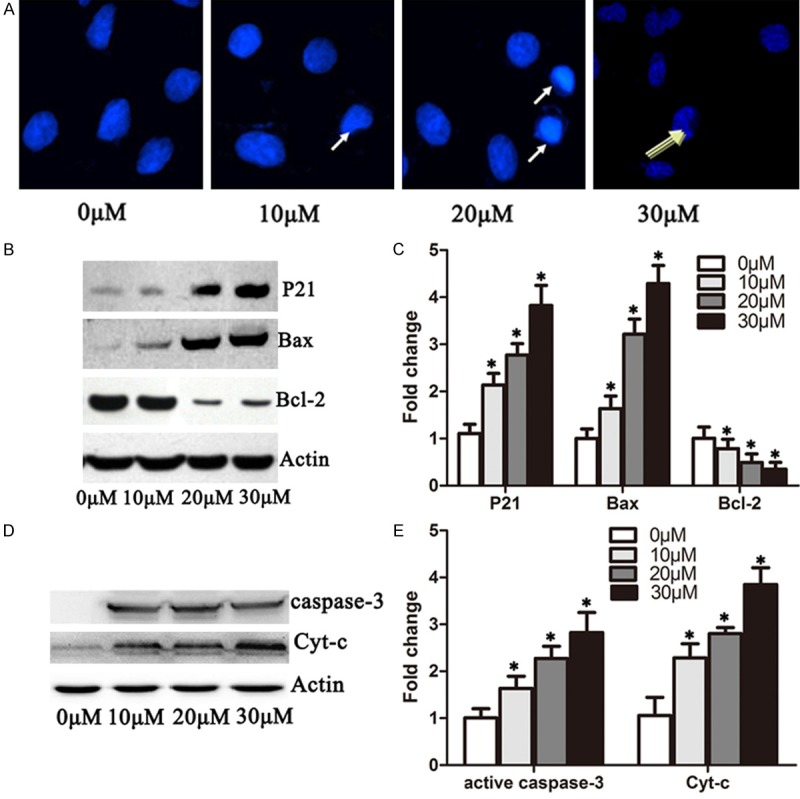
Figure 3.

DHA induced apoptosis in A549 cells by increasing p21, Bax, cytochrome-c and active caspase 3 protein level and inhibiting Bcl-2. A. Immufluorescence microscopy analysis of 549 cells treated with 0 μM, 10 μM, 20 μM and 30 μM DHA for 72 hours. Cells were stained with propidium iodide. The condensed nucleus and cleaved nucleus were enhanced by 20 μM and 20 μM DHA as the arrow indicates. B. DHA cells were treated with 0 μM, 10 μM, 20 μM and 30 μM of DHA 48 hours, respectively. Western blots were performed to detect the protein levels of p21, Bax, Bcl-2 and actin. C. Quantification of p21, Bax and Bcl-2 protein levels from immunblots in B. The protein levels of p21, Bax and Bcl-2 were normalized to beta-actin. D. A549 cells were treated as in B. Western blots were performed to detect the protein levels of cytochrome-c and active-Caspase 3. E. Quantification of cytochrome-c and active-Caspase 3 levels from immunoblots as in D. The protein levels of cytochrome-c and active-Caspase3 were normalized to beta-actin. *P < 0.05.
