Figure 4.

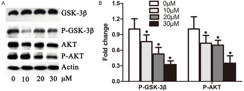
DHA reduced the activity of the AKT/GSK-3β pathway. A. A549 cells were treated with 0 μM, 10 μM, 20 μM and 30 μM DHA for 48 hours, respectively. Total proteins were extracted for the immunoblotting of AKT, p-AKT, GSK-3beta, p-GSK-3beta and actin. B. Quantification of p-AKT and p-GSK-3beta levels from immunoblots in A. The protein levels of p-AKT and p-GSK-3beta were normalized to beta-actin. *P < 0.05.
