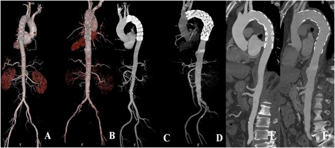
Fig 1. 2D and 3D reconstructions with images generated using low-iodine and high-iodine groups.
Volume rendering, maximum-intensity projection and multiplanar reformation images (A-F) show endovascular repair of aortic dissection (A, C, E) and aneurysm (B, D, F) with stent graft placed just below the left subclavian artery. A, C and E represent images acquired with the low-iodine protocol, while B, D and F are images generated with the high-iodine protocol. There is no difference in the visualization of stent graft and aortic branches between the two groups.

