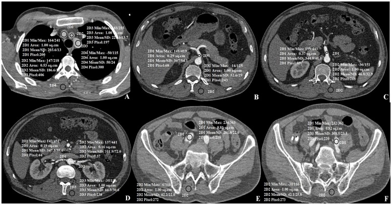
Fig 3. Axial images of aortic branches and segments with paraspinal muscle.
A) Lumen of the Brachiocephalic trunk (2D1), the Left common carotid artery (2D2) and the Left subclavian artery (2D3), B) Celiac trunk, C) the superior mesenteric artery, D) the right renal artery (2D1) and the left renal artery (2D2), E) the right common iliac artery, F) the left common iliac artery. Mean attenuation values with standard deviations are shown in the images.

