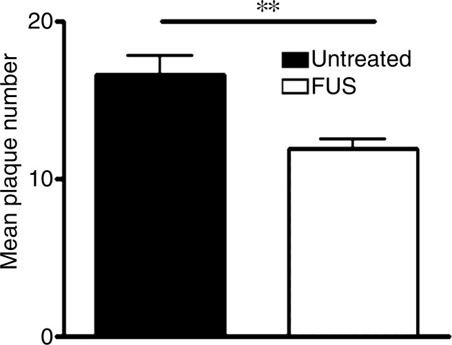
Figure 3d:
MR imaging–guided focused ultrasound (FUS) reduces plaque pathologic abnormalilties in the TgCRND8 brain. Plaques were identified by using the 6F3d antibody and were quantified by using ImageJ analysis. (a, b) Representative 10× images of the plaque pathologic abnormalities in untreated TgCRND8 mice (a) and MR imaging–guided focused ultrasound –treated TgCRND8 mice (b). (c) Bar graph shows that mean plaque size in the hippocampus of untreated TgCRND8 mice was 352 µm2 ± 38 which was reduced by 20% to 279 µm2 ± 16 after MR imaging–guided focused ultrasound (P < .01). (d) Bar graph shows that the total number of plaques in the hippocampus of untreated TgCRND8 mice was a mean of 17 ± 3 and was reduced to a mean of 12 ± 1 after MR imaging–guided focused ultrasound treatment, representing a reduction of 19% (P < .01). In c and d, bars represent means, and error bars represent standard errors of the mean, with n = 6–8 per group. ** = P < .01.

