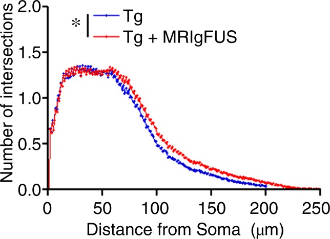
Figure 4e:
MR imaging–guided focused ultrasound (MRIgFUS) treatment increases DCX-positive number of cells and branching. (a) Images from DCX immunocytochemical analysis was performed in four equally spaced sections from all animals. Representative 10× images of the newborn neurons in the hippocampus are provided for each treatment group. (b) Bar graph shows that the number of DCX-positive cell bodies increased by 188% in non-Tg mice (P < .05) and by 252% in TgCRND8 mice treated with MR imaging–guided focused ultrasound (P < .05). (c) Bar graph shows that the total dendrite path length was increased by 227% in non-Tg mice (P < .05) and by 332% in TgCRND8 mice (P < .01) treated with MR imaging–guided focused ultrasound. (d) Graph depicts that the number of intersections of a dendrite with the concentric circles, which describes the relative amount of dendrite branching, showed that MR imaging–guided focused ultrasound treatment increased dendritic branching in non-Tg mice at distant radii (P < .05). (e) Graph depicts that the dendritic branching was also increased in TgCRND8 mice following repeated treatment with MR imaging–guided focused ultrasound (P < .05). In b and c, bars represent means, and error bars represent standard errors of the mean, with n = 6–8 per group. * = P < .05, ** = P < .01.

