Figure 2:
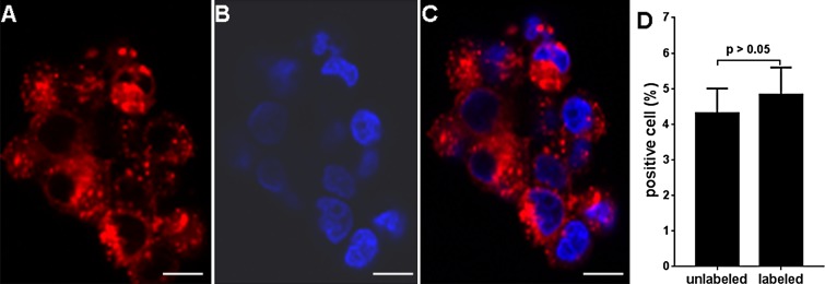
A–C, Images from fluorescence microscopy of iron-labeled dendritic cell vaccines and, D, results of trypan blue assessment of labeled dendritic cells. A, Texas Red particle accumulation is observed in cytoplasm while, B, DAPI-stained nuclei were blue, with coregistration in, C, a merged image. D, Bar graph shows that there was no significant difference between trypan blue viability indexes for labeled and those for unlabeled dendritic cells. Scale bars for A, B, and C = 10 μm.

