
Fig. 3.
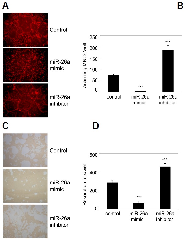
miR-26a attenuates actin ring formation and bone resorption. (A, B) BMMs were cultured for 2 days in the presence of M-CSF and RANKL. Two-day-old pre-OCs were transfected with a miR-26a mimic, miR-26a inhibitor, or negative control as indicated. Transfected cells were further cultured in the presence of M-CSF and RANKL. (A) Cells were fixed and stained with phalloidin. (B) Numbers of MNCs containing an actin ring were counted. Data represent means ± SD of triplicate samples. ***p < 0.001 vs. control. Results are representative of at least three independent sets of similar experiments. (C–D) BMMs were cultured on Osteo Assay plates for 2 days in the presence of M-CSF and RANKL. Two-dayold pre-OCs were transfected with a miR-26a mimic, miR-26a inhibitor, or negative control as indicated. Transfected cells were further cultured for 36 h in presence with M-CSF and RANKL. (C) Resorption lacunae were visualized by bright-field microscopy. (D) Numbers of resorption pits were counted. Data represent means ± SD of triplicate samples. ***p < 0.001 vs. control. Results are representative of at least three independent sets of similar experiments.