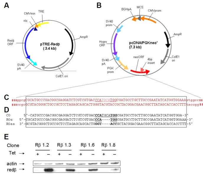
Fig. 1.

Reagents used for Redβ expres sion and ssOR assays. (A) Structure of Redβ expression plasmid. (B) Structure of plasmid carrying the neo* target for gene correction. (C) Sequence surrounding the 4 bp insertion (bold underlined) in neo*. (D) Sequence of control (CO) and repair (RO) ssOs used in ssOR assays. (E) Immunoblot analysis of Redβ expression in hygroR clones isolated after cotransfection of HT1080 cells with the plasmids shown in (A) and (B). Clones were expanded and grown continually with tetracycline (Tet) in the growth medium (+), or for 3 days after the removal of tetracycline (−), before analysis.
