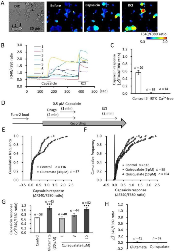
Figure 2.

Effects of glutamatergic drug perfusion on capsaicin-induced elevation of intracellular calcium in primary cultured DRG neurons. (A) Representative images of F340/F380 ratio before and after the perfusion of capsaicin (0.5 μM) and KCl (50 mM) using Fra-2 AM dye. (B) Time course of F340/F380 ratio in seven independent cells from panel A. Horizontal bars represent periods of capsaicin and KCl perfusion. (C) Effects of 5′-IRTX and calcium-free solution on capsaicin-induced elevation of intracellular calcium in DRG neurons. (D) Experimental design for the recording of capsaicin-induced calcium elevation during glutamatergic drug perfusion. (E, F) Cumulative frequency graph showing changes in the F340/F380 ratio induced by capsaicin in the presence of glutamate and quisqualate. (G) Quantitative data showing changes in the F340/F380 ratio induced by capsaicin in the presence of glutamate and quisqualate. Data represents average of 50% of neurons, those with the largest capsaicin-induced calcium elevation. (H) Changes in intracellular calcium levels during glutamate or quisqualate perfusion. Data presented as mean ± SEM. ***P < 0.0001, significantly different from control.
