Figure 5.
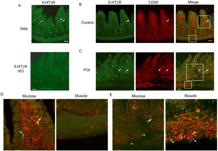
Double-staining for 5-HT3R and CD68-positive macrophages in ileum of mice. (A) Examination of specificity of anti-5-HT3 receptor antibody. Upper panel or lower panel shows immunohistochemistry by anti-5-HT3 receptor antibody in ileum of wild-type mice or 5-HT3A receptor null mice respectively. Each picture shows a typical result from three independent experiments using two mice. (B) Double-staining of 5-HT3 receptor and CD68 in the ileum of control mice and POI mice. Arrow shows double-positive cells. Typical results from four independent experiments are shown. (D and E) Higher magnification pictures in both mucosal and muscle layer of ileum in control mice (D) or POI mice (E). Each picture was magnified from square area of (B) and (C). Arrow head and arrow indicate double-positive cells. Green or red stain indicates 5-HT3 receptors or CD68 respectively. Scale bar shows 50 μm.

