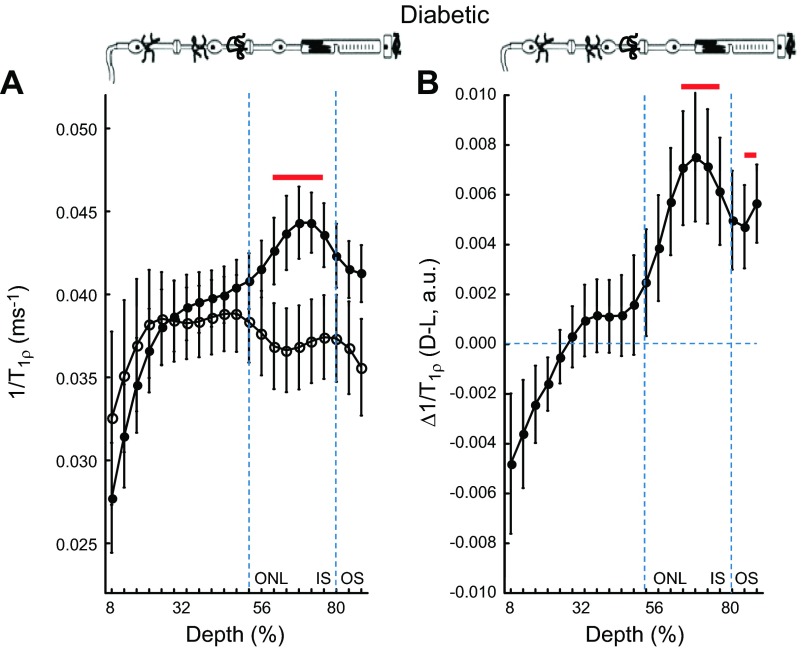
Figure 4.
1/T1ρ MRI measurements predict normal light-evoked tet-ARR1 translocation and visual cycle activity in a model of diabetic retinopathy. A) Layer-specific analysis of mean group central retinal 1/T1ρ profiles during darkness (solid circle, D) and light (open circle, L) in manganese-treated diabetic mice. B) Paired-difference profile between dark and light 1/T1ρ (i.e., Δ1/T1ρ) profiles in diabetic mice using the data in (A). Data are presented using the conventions in Fig. 1. In the outer retina, 1/T1ρ data indicates normal responses, suggesting that diabetes has little effect on light-dependent ARR1 translocation—and thus visual cycle activity—at this duration of disease; this prediction was confirmed by the data in Fig. 5. We also note that the Δ1/T1ρ profile in the inner retina of the diabetic mice was unresponsive to light. Our data suggest (section C) that 1/T1ρ profile in the inner retina is an independent measure of functional change in retinal blood volume (i.e., autoregulation); this complements, and is not necessarily related to, ARR1 translocation evaluation. We previously established impaired autoregulation in diabetic mice (27), and the present data provide additional support for this phenotype.

