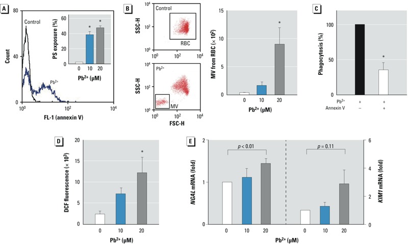
Figure 2.

Role of Pb2+-induced erythrophagocytosis in renal tubular damage. (A,B) Erythrocytes (red blood cells; RBCs) were incubated with Pb2+ for 1 hr at 37°C, and flow cytometric analysis was used to determine externalization of phosphatidylserine (PS) in the outer membrane (A) or generation of microvesicles (MVs) from erythrocytes (B); representative histograms and quantified bar graphs are shown. (C) Pb2+-induced erythrophagocytosis in HK-2 cells was reversed by the addition of purified annex V, which masked externalized PS in erythrocytes. (D,E) Renal tubular damage determined after co-incubation of HK-2 cells with Pb2+-treated erythrocytes for 4 hr at 37°C. Generation of ROS was measured by dichlorofluorescein fluorescence in HK-2 cells by flow cytometry (D), and mRNA levels of the nephrotoxic biomarkers NGAL and KIM1 were detected in HK-2 cells by qRT-PCR (E). Data were analyzed by one-way ANOVA followed by Duncan’s multiple range test (A,B,D) or Student’s t-test (C,E) to determine statistical significance. Values shown are the mean ± SE of more than three independent experiments. *p < 0.05 compared with the corresponding control.
