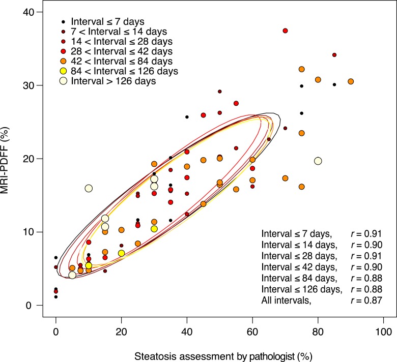
Figure 3:
Scatterplot of MR imaging–estimated PDFF versus near-continuous steatosis score (mean of two readings) is shown, according to the time interval between MR imaging and liver biopsy. No significant effect of the time interval between MR imaging and biopsy was observed on the relationship between MR imaging–estimated PDFF and steatosis.

