
Figure 2.
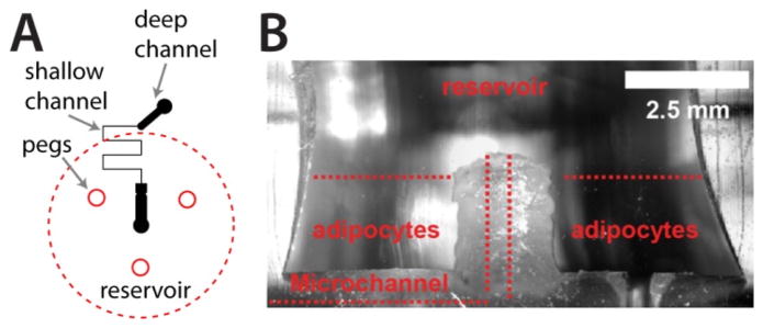
Macro-to-micro interface for primary tissue culture and sampling. A) The microfluidic channel layout is depicted in black, with deep (156 μm) and shallow (15 μm) channels included. Insert peg locations and reservoir placement are shown in red. B) A cross section of a typical PDMS interface, highlighting the “moat” region for culture of primary adipocytes that addresses cell buoyancy issues and allows passive secretion sampling into the microchannel.