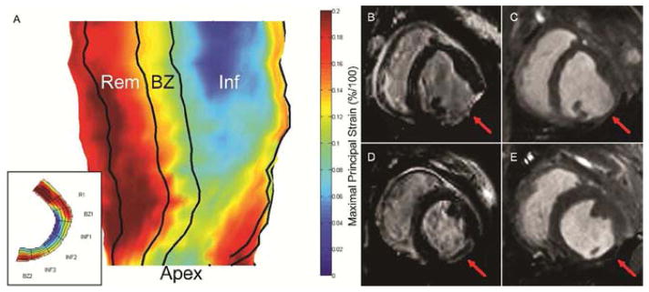
Figure 1.

(A) 3D radial strain map generated from manually segmented mid-myocardial contours. 2D short-axis strain maps (inset) are first generated for each myocardial slice from apex to base. Infarct, borderzone, and remote boundaries are then identified. Inf – Infarct region, BZ – Borderzone region, Rem – Remote region, Apex – LV apex. (B–E) Late gadolinium enhanced and cine images of 4wk untreated control (B & C) and 4wk Radiesse® treated (D & E) infarcts at end diastole. Red arrow denotes infarct area. Diffuse Radiesse® deposits are visible within the infarct scar as a black void.
