Figure 4.
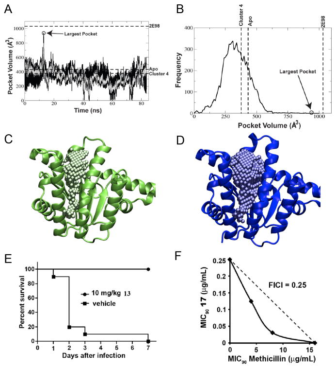
Molecular dynamics as a route to drug lead discovery. A–D, MD results for E. coli UPPS. A, volume of the binding pocket along the MD trajectory of E. coli undecaprenyl diphosphate synthase (UPPS). The black line shows data taken every 10 ps, the over-layed gray line is the average over every 100 ps; B, frequency at which different volumes of the pocket are sampled; C, the apo crystal structure with 1 Å spheres filling the active site pocket; D, a bisphosphonate-bound crystal structure with 1 Å spheres filling the active site pocket. Note the significantly larger pocket size in the bisphosphonate-bound structure when compared to the apo crystal structure. The MD-based structures provide the best correlation between experimental IC50 values and docking scores. E, activity of 13 (bisamidine, BPH-1358) in a mouse model of S. aureus (USA200) infection; F, in vitro synergy showing isobologram for BPH-1358 + methicillin inhibition of S. aureus (USA300) cell growth, FICI = 0.25. A to D are reprinted with permission from reference [40]. E and F are reprinted with permission from reference [34].

