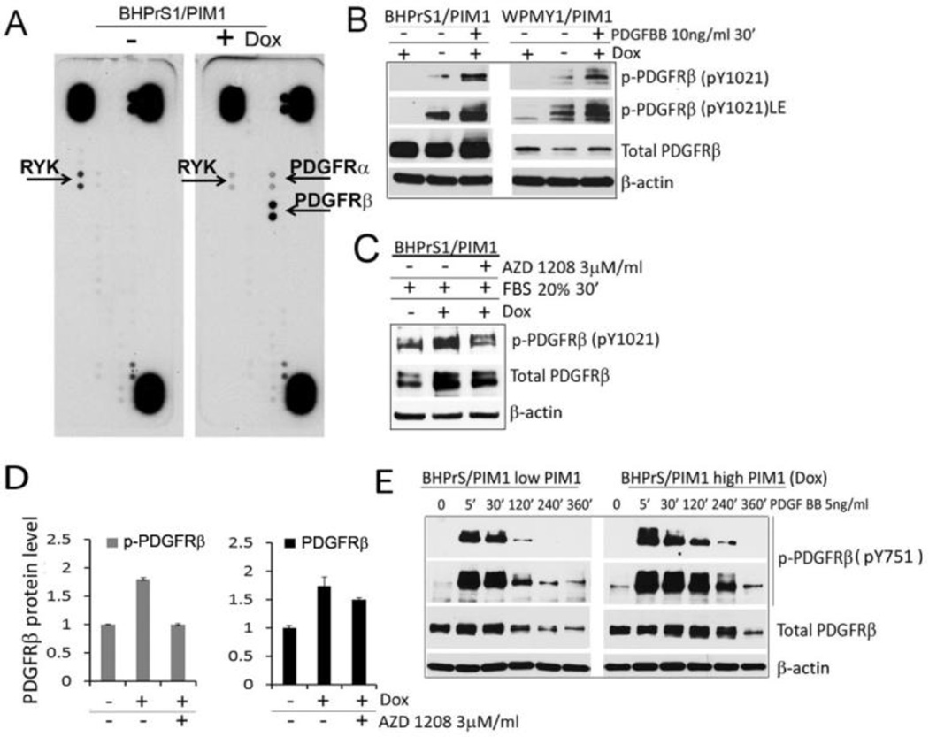
Figure 4. PIM1 kinase expression leads to increases PDGFR level and phosphorylation.
(A) BHPrS1stable pools expressing inducible PIM1 kinase were incubated with or without Dox (20 ng/ml) for 48 hours, then lysed and 300µg of total proteins were assayed by the human phospho-RTK array (R&D Biosystem). Arrows indicate RYK and PDGFR kinases. (B) BHPrS1/PIM1 and WPMY1/PIM1 cells with or without PIM1 induction were incubated in a serum free media overnight, and then treated with PDGFRβ ligand (PDGFBB) for 30 min. Cell lysates were analyzed by Western blotting for the indicated proteins. (C) BHPrS1/PIM1 cells with and without PIM1 induction by Dox were incubated in serum free media overnight in the presence of DMSO (vehicle control) or 3µM AZD1208, the cells were then stimulated with 20% FBS for 30 min and assayed by Western blotting with the indicated antibodies. (D) Spot densitometry analysis was performed for PDGFRβ protein levels seen in C and intensity was normalized to the β-actin loading control. (E) BHPrS1/PIM1 cells with and without PIM1 induction by Dox were incubated in serum free media overnight and PDGFRβ activation was induced by PDGFBB ligand for indicated time period.

