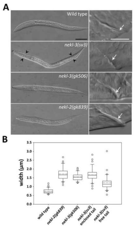
Fig. 3. Unshed cuticles of the nekl-2 and nekl-3 mutants.
(A) DIC images of arrested nekl-3(gk506) and nekl-2(gk893) homozygotes compared with a wild-type L2 larva and a nekl-3(sv3) homozygote arrested at L2. In nekl-3(gk506) and nekl-2(gk893) homozygotes, the cuticle always encloses the entire larva, whereas nekl-3(sv3) mutants typically shed the cuticle in the head and tail regions (black arrowheads). White arrows in right panels indicate corresponding rectums, which are thicker in nekl-3(gk506) and nekl-2(gk893) homozygotes because of a double layer of cuticle. Scale bar for left panels = 50 µm; scale bar for right panels = 10 µm. (B) Box-and-whisker plot showing the rectal width of mutants. The box represents the middle 50% of the data points that are the closest to the median value (black line); the dashed red line represents the mean value. Dots are outliers that differ by ≥1.5-fold from the upper or lower quartiles; whiskers encompass values between the outliers (dots) and the upper and lower quartiles (top and bottom of box). sv3 homozygotes with free tails are somewhat variable in rectal widths but are closer to wild type than are nekl-2(gk506) or nekl-3(gk893) deletion mutants. sv3 homozygotes with enclosed tails have rectal widths that are indistinguishable from deletion mutants. n ≥ 30 for each genotype. For raw data and statistical analyses, see Supplementary File 1.

