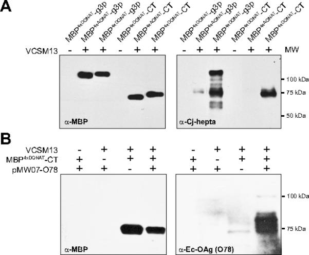
Figure 2. Generation of phage-displayed glycan library members.

(A) Western blot analysis of phage preparations (containing 4×10¹⁰ glycophage particles) derived from E. coli TG1 ΔwaaL cells co-transformed with pMW07pglΔB and one of the following phagemids: pBAD-MBP4xDQNAT-g3p::PglB, pBAD-MBP4xAQNAT-g3p::PglB, pBADMBP4xDQNAT-CT::PglB or pBAD-MBP4xAQNAT-CT::PglB. (B) Western blot analysis of phage preparations (containing 6×10¹⁰ glycophage particles) derived from E. coli TG1 ΔwaaL cells co-transformed with phagemid pBAD-MBP4xDQNAT-CT::PglB and pMW07-O78. Phage preparation involved addition (+) or omission (-) of VCSM13 helper phage. Additional controls involved omitting either the phagemid or the glycan biosynthesis pathway. Blots were probed with anti-MBP, hR6 serum (anti-Cj-hepta), or anti-Ec-OAg (O78) as indicated. Molecular weight (MW) markers are indicated at right.
