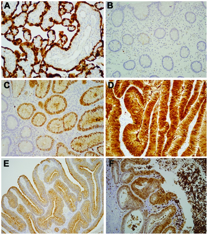
Figure 3.
Immunohistochemical detection of S100P protein expression in placenta and colon tissue. (A) Trophoblasts lining the placental chorionic villi demonstrated uniform and strong staining (magnification, ×200). (B) Normal colonic mucosa showed negative staining (magnification, ×200). (C) Glands with low-grade dysplasia showed heterogeneous nuclear staining and weak cytoplasmic staining (magnification, ×200). (D) Strong nuclear and moderate cytoplasmic staining in glands with high-grade dysplasia (magnification, ×400). (E) Strong staining in the apical region of cytoplasm without nuclear expression (magnification, ×100). (F) Neutrophils forming abscess around invasive cancer displayed very strong immunostaining (magnification, ×200).

