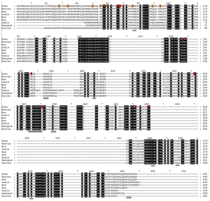
Figure 3.
Positive selection sites in planar structure. Amino acid residuals in orange background belong to positive selection sites detected using the site model. Amino acid residues in red background belong to positive selection sites detected using the branch model and branch-site model. #, protein kinase C phosphorylation site; &, casein kinase II phosphorylation site; @, serine active site.

