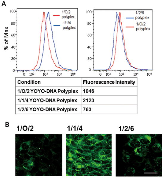
Figure 9. Quantification of DNA uptake in MCF-7/Adr cell line.
(A) Flow cytometry quantified P(SiDAAr)5P3 polyplexes' DNA uptake levels in MCF-7/Adr cell line. 1/1/4 polyplexes showed over 2-fold higher DNA uptake than 1/O/2 polyplexes. The level dropped in 1/2/6 polyplexes due to polymer masking. (B) Confocal microscopy visualized YOYO-1-DNA's cellular internalization. 1/1/4 polyplexes showed a stronger DNA internalization than 1/O/2 polyplexes. Fluorescence intensity dropped in 1/2/6 polyplexes, confirming the flow cytometry results. Images were captured using 80× objective lens. Bar=50 μm.

