
Figure 6.
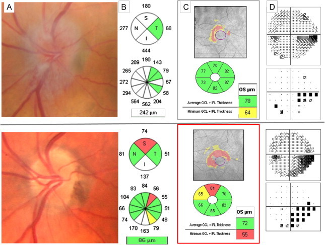
Left anterior ischemic optic neuropathy. Top row: One week after acute visual acuity loss. (A) Optic nerve swelling causes RNFL thickening that prevent evaluating if there is or not associated axonal loss (B). However GCIPL analysis (C) shows an abnormal thinning that correlates with the inferior VF defect (D). Bottom row: two months after the acute episode. Superior optic disk pallor (A), superior RNFL quadrant thinning (B) and GCIPL thinning (C) with the corresponding inferior VF defect (D).