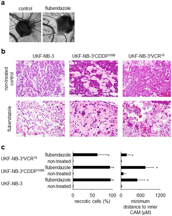
Figure 6. In vivo-effects of flubendazole in the chick chorioallantoic membrane (CAM) model.
Flubendazole was applied as water-soluble (2-hydroxypropyl)-β-cyclodextrin complex. (a) Representative photographs demonstrating the effects of flubendazole (20 μg/mL) on vessel formation in the CAM compared to a vehicle-treated control; (b) haematoxylin/eosin staining of representative CAMs demonstrating the effects of flubendazole (2 μg, applied as (2-hydroxypropyl)-β-cyclodextrin complex in 100 μL 0.9% NaCl solution) (original magnification: 20-fold). Flubendazole-treated tumours grow in a less extensive manner and present with large necrotic areas. (c) Quantification of necrosis formation and cancer cell invasion in the CAM was studied in UKF-NB-3 (5 non-treated tumours, 8 flubendazole-treated tumours), UKF-NB-3rCDDP1000 (4 non-treated tumours, 8 flubendazole-treated tumours), and UKF-NB-3rVCR10 (9 non-treated tumours, 9 flubendazole-treated tumours). To measure cancer cell invasion, the minimum distance of the cancer cell invasion front to the inner CAM border was determined. Higher values indicate a greater distance to the inner membrane and, therefore, lower invasion. * P < 0.05 relative to non-treated control.

