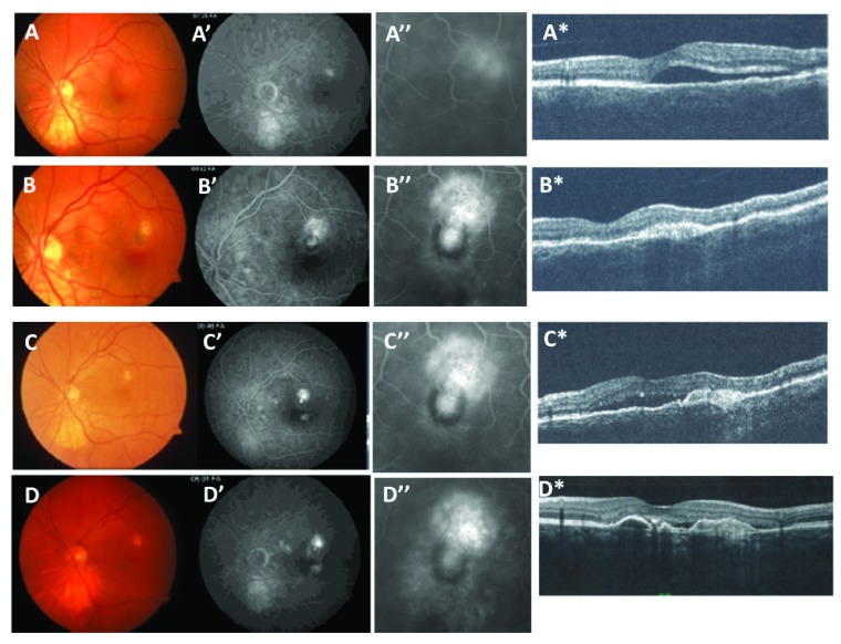
Figure 1. Partial pigment ring.
Fundus photograph ( A–D), fluorescein angiogram (FA) mid-phase ( A’–C’), magnified FA ( A”–D”) and OCT ( A*–D*) images from a 58 year old female patient presenting with worsening visual acuity to 20/30 and metamorphopsia. Figures 1A-A*) Images taken prior to treatment show a peri-foveal fluorescein leakage with serous detachment due to CNV. The patient was treated with thermal laser to ablate the CNV lesion. Figures 1B-B*) After 6 months, CNV recurred in the supero-temporal aspect of the laser scar. The recurrence was treated with combined therapy of 2 PDT and 4 bevacizumab injections applied over a 6 month period. Figure 1C-C*) After remaining stable for 9 months without treatment, the CNV again recurred in the direction of the thermal laser scar but not in the direction of the partial pigment ring. This recurrence was treated with serial anti-VEGF antibody injections and then remained stable after 42 months with anti-VEGF therapy as shown in Figure 1D-D*.

