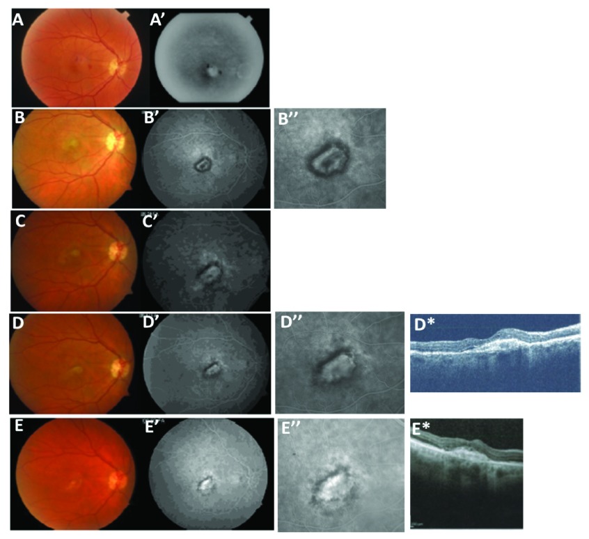
Figure 2. Recurrent pigment capping.
( Figures 1A-A’) shows a 71 year old female patient who presented with decreasing vision over 1 week with fundus photography and angiography indicating perifoveal CNV and a small amount of hemorrhage. Figures 2B-B”) After 5 visudyne PDT treatments over a 13 month period, a complete ring of pigment formed to surround the CNV and treatment was withheld. Note the reduced fluorescein leakage. Figures 2C-C’) The treatment interval was extended to 20 months after which symptoms and a small infero-temporal area of fluorescein leakage recurred. Anti-VEGF treatments with bevacizumab and pegaptanib were initiated and after 7 treatments over a 31 month period, a pigment ring re-formed around the CNV with elimination of the infero-temporal leakage as shown in Figures 2D-D*. After this, the treatment interval was again extended to 21 months without treatment during which the lesion remained stable as shown in Figures 2E-E*.

