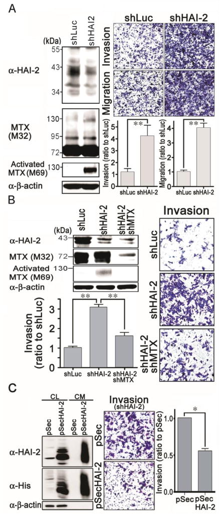
Figure 5.
Effect of HAI-2 knockdown on matriptase, PCa cell invasion and migration, as well as the effects of matriptase knockdown and recombinant HAI-2 proteins on HAI-2-silencing-induced cancer cell invasion. (A) Stable pools of HAI-2 knockdown CWR22Rv1 cells were established by G418 selection after the cells were transfected with shHAI-2 plasmids. The effects of HAI-2 silence on matriptase, cell invasion and migration were analyzed using the same methods as in Figure 4B and 4C. (B) Examination of the involvement of matriptase in increased cell migration and invasion by HAI-2 knockdown. HAI-2-knockdown CWR22Rv1 cells were infected with shMTX viral particles for 24 h and selected for three days. Cell lysates were analyzed by immunoblotting with anti-HAI-2, anti-matriptase (M32), anti-activated matriptase (M69) and anti-β-actin Abs. Infected cells were seeded at a number of 4 × 10⁵ cells per chamber and cultured for 20 h. Invaded cells were stained, photographed, and measured as described in Figure 4C. (C) Effect of recombinant HAI-2 proteins on the HAI-2-knockdown-induced cancer cell invasion. The DNA fragment encoding the extracellular region of HAI-2 was cloned into a secretory vector pSecTag2. HEK293T cells were transfected with the plasmid or vector alone and the stable pools were selected by G418. The stable pools were seeded at a density of 3 × 10⁶ cells in a 6-cm plate within a regular culture medium. The next day, the culture media were refreshed with serum-free RPMI1640 media. Twenty four hours after the refreshment, cell lysates (CL) and the conditioned media (CM) were collected. The protein concentrations in the cell lysates were determined by Protein Assays. Equal amounts of cell lysates and the conditioned media from the same cell numbers by normalizing to the whole cell lysates were used for SDS-PAGE and immunoblot analysis with anti-HAI-2 and anti-His Abs. The conditioned media were used for the cell invasion assays by adding to the upper and lower chambers. In the lower chambers, 10% FBS were supplemented as chemoattractant. The cell invasion was analyzed as the same methods as described previously. *, P< 0.05; **, P<0.01.

