
Figure 2.
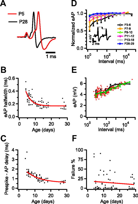
Developmental changes in timing and reliability of synaptic transmission. A, The first complex waveform in the middle panels of Figure 1, A and C, are aligned on the prespike and scaled to the same peak amplitude to illustrate the difference in the time course of the complex waveform at P5 and at P28. B, Developmental changes in eAP half-width. The solid line is an exponential fit with time constant 4 d. C, Developmental changes in latency between prespike and eAP. The solid line is an exponential fit on the data starting at P5, with time constant 8 d. D, Depression of eAP size as a function of eAP interval. eAP sizes from individual recordings (n = 57) were log binned and normalized to the average of the 3 bins with the longest intervals before averaging within the different age groups. Inset shows example of depression of eAP size at short intervals in a P5 rat. E, Relation between eAP size and log interval. Green trace shows the binned average with SD. Black trace shows model fit with single recovery time constant of 320 ms. F, Percentage of subthreshold eEPSPs per cell as a function of age. Red line shows linear regression (r = −0.1; p > 0.4).