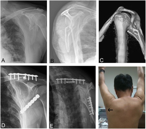
Figure 1.

Course of double fixation. Anteroposterior (A) and lateral (B) radiographs of the patient with ipsilateral fracture of the clavicle and the scapula neck before the treatment. The glenopolar angle was 3°; three-dimensional computerized tomography image showed the scapular neck fracture angulation was up to 43° (C); post-operative follow-up radiographs revealed the glenopolar angle was 36° (D, E); the post-operative photograph showed the operated shoulder (F) and a straight incision over the glenoid neck to access minimal dissection between the teres minor and the infraspinatus (arrow).
