Fig. 6.
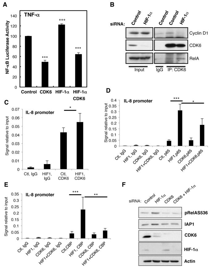
HIF de-repression of NF-κB activity requires CDK6. (A) HeLa-κB cells were co-transfected with the indicated siRNA for 48 h. Cells were treated with TNF-α for 6 h, and the luciferase activity was measured. The means+s.d. were determined from three independent experiments. Student’s t-test analysis was performed *P≤0.05, **P≤0.01, ***P≤0.001. (B) HeLa cells were transfected with control or HIF-1α siRNA oligonucleotides for 48 h before lysis. To immunoprecipitate CDK6, 200 μg of protein was used. Normal rabbit IgG was used as a control. Immunoprecipitated complexes were analysed by western blotting using the indicated antibodies. Inputs correspond to 10% of material. (C) HeLa cells were transfected with control (Ctl) and HIF-1α (HIF1) siRNA oligonucleotides for 48 h before treatment with 10 ng/ml TNF-α for 1 h. Cells were fixed and lysed, and chromatin immunoprecipitation assays were performed using the indicated antibodies. Purified DNA was analysed by using qPCR with primers for the IL-8 promoter. The graph depicts means+s.d. from three independent experiments. Student’s t-test analysis was performed *P≤0.05, **P≤0.01, ***P≤0.001. (D,E) HeLa cells were transfected with control (Ctl), HIF-1α (HIF1), CDK6 (CDK6) or HIF-1α and CDK6 (HIF1+CDK6) siRNA oligonucleotides for 48 h before treatment with 10 ng/ml TNF-α for 1 h. Cells were processed and analysed as in C. The graph depicts means+s.d. from three independent experiments. Student’s t-test analysis was performed *P≤0.05, **P≤0.01, ***P≤0.001. (F) HeLa cells were transfected with control, HIF-1α, CDK6 or HIF-1α and CDK6 siRNA oligonucleotides for 48 h before lysis. Whole-cell lysates were analysed by western blotting for the indicated proteins. See also supplementary material Fig. S5.

