Figure 2.
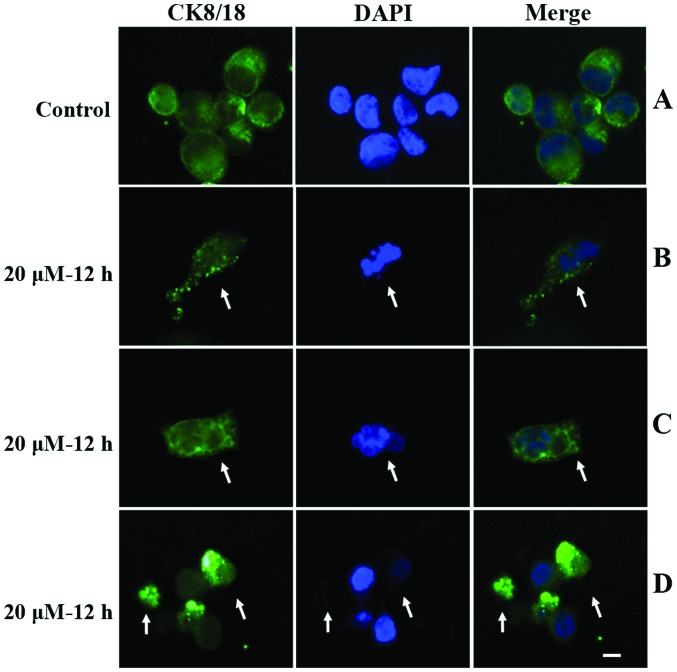
Fluorescein-labeled anti-CK8/18 antibody was used to detect apoptotic cells. (A) Control cells showed regular nuclear DAPI staining with uniform distribution of cytoplasmic CK8/18 staining. Apoptotic cells with fragmented nuclei exhibited (B) punctate or (C) bubbly CK8/18 distribution in cytosol. (D) Apoptotic cells with a split nucleus and weak DAPI staining also exhibited punctate or aggregated CK8/18 staining in the cytosol. Arrows indicate apoptotic cells. Scale bar, 10 μm. CK, cytokeratin; DAPI, 4′,6-diamidine-2′-phenylindole dihydrochloride.

