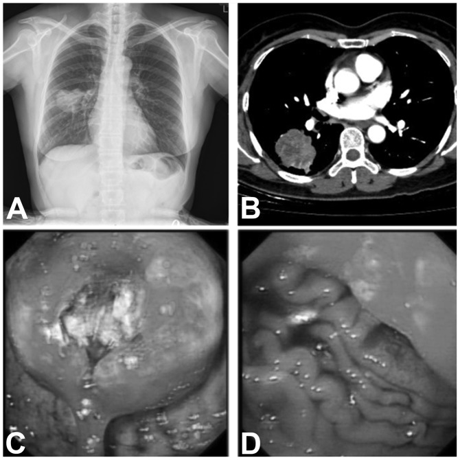
Figure 2.
(A) Chest X-ray revealing an abnormal mass shadow in the right lower lobe. (B) Computed tomography scan of the chest demonstrating an irregular mass measuring 39×48 mm in size, with a scallop-shaped contour and focal enhancement. (C and D) Gastroscopy images revealing a mass, ~4×4 cm in size, in the fundus of the stomach, invading the cardia, with a deep ulcer in the center.

