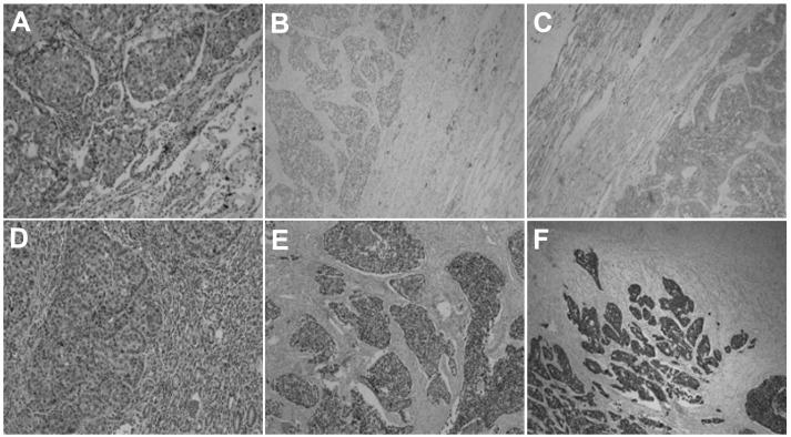
Figure 3.
(A) Cancer tissue revealing the atypical nest and mesh shape of the cell arrangement (stain, hematoxylin and eosin; magnification, ×100). The positive immunohistochemical staining for (B) TTF-1 and (C) CK-7 indicates that the cancer originated from the lung. (D) HE staining of the gastric mass tissues (magnification, ×100). The cancer tissue exhibits similar HE morphology to lung adenocarcinoma, and there are clear boundaries between the cancer tissue and the normal gastric gland. Positive immunohistochemical staining for (E) TTF-1 and (F) CK-7 which is the same result as found in lung adenocarcinoma. TTF-1, thyroid transcription factor-1; CK, cytokeratin; HE, hematoxylin and eosin.

