Figure 1.

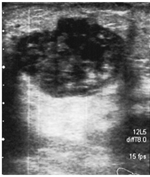
Ultrasonography of the tumor mass. The image reveals the presence of a hypoechoic solid neoformation with jagged edges and a maximum diameter of ~3 cm.

Ultrasonography of the tumor mass. The image reveals the presence of a hypoechoic solid neoformation with jagged edges and a maximum diameter of ~3 cm.