Figure 11.

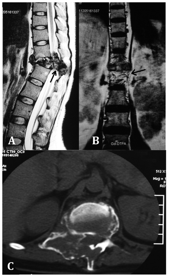
Case two. (A) Sagittal, (B) coronal and (C) transverse plane magnetic resonance imaging showing the additional mass in the spinal canal and posterior column of the T12 vertebra.

Case two. (A) Sagittal, (B) coronal and (C) transverse plane magnetic resonance imaging showing the additional mass in the spinal canal and posterior column of the T12 vertebra.