
Figure 2.
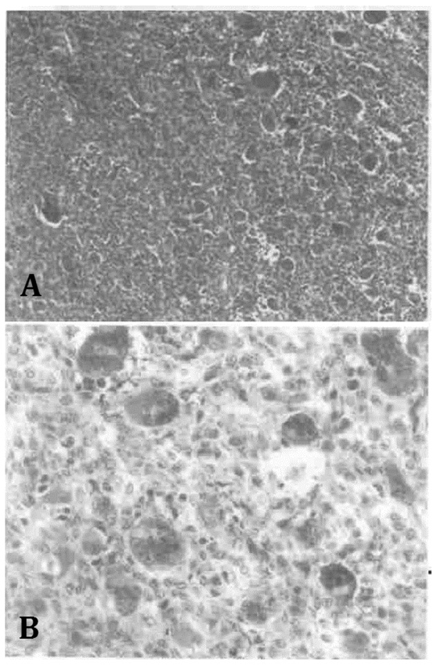
Case one (magnification, ×100). Immunohistochemical staining for the (A) pre-operative fine-needle biopsy and (B) post-operative resection specimens showed the tissues to be AE1/AE3(−), cluster of differentiation 68(+), p53(+) and S-100(+), with a Ki-67 of 20%. The histopathological examinations of the lesion established a diagnosis of a giant cell tumor of the bone.