Figure 3.

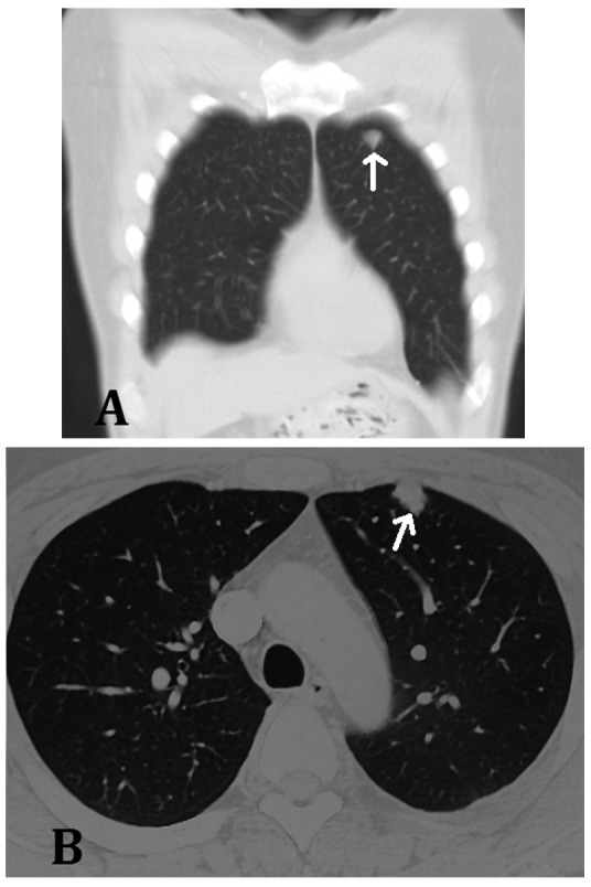
Case one. (A) Coronal and (B) transverse computed tomography scans of the chest, performed four months after the first surgery at the follow-up visit, showing a nodule with clear borders in the anterior upper left lobe of the lung.

Case one. (A) Coronal and (B) transverse computed tomography scans of the chest, performed four months after the first surgery at the follow-up visit, showing a nodule with clear borders in the anterior upper left lobe of the lung.