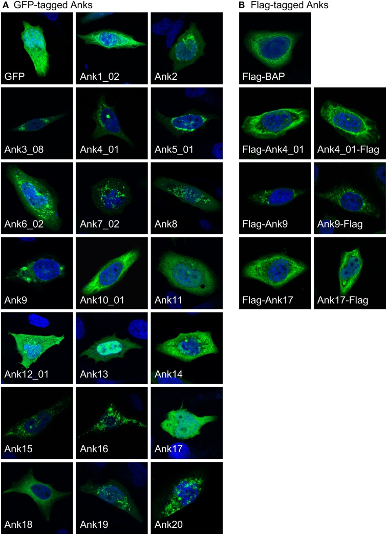
Figure 6.
Ectopically expressed O. tsutsugamushi Anks exhibit diverse subcellular localization patterns. (A) HeLa cells expressing GFP alone or the indicated Ank proteins N-terminally fused to GFP were screened with GFP antibody, and visualized using confocal microscopy. (B) Subcellular localization patterns of ectopically expressed Anks are not affected by the fusion tag itself or tag placement. HeLa cells expressing the indicated Anks as N-terminally (Flag-Ank) or C-terminally Flag-tagged (Ank-Flag) fusion proteins or Flag-BAP were screened with Flag tag antibody and examined by confocal microscopy. (A,B) HeLa cell nuclei were stained with DAPI (blue). Representative images from 2 to 4 experiments performed per each ectopically expressed Ank are presented.

