
Figure 7.
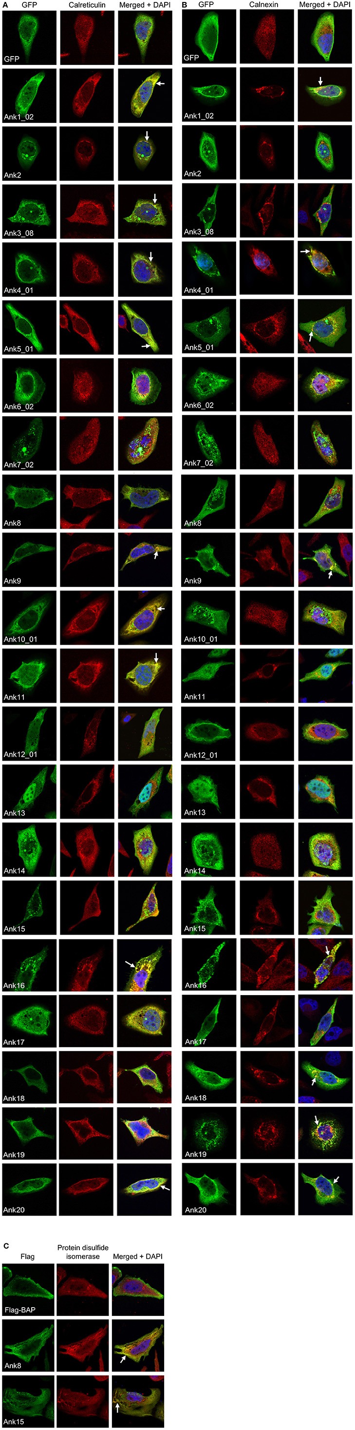
Multiple ectopically expressed O. tsutsugamushi Anks localize to the endoplasmic reticulum. HeLa cells expressing GFP alone or GFP-Anks were screened with GFP antibody, and cells expressing Flag-BAP or Flag-Anks were screened with Flag antibody. Additionally, cells were stained with antibody against either of the ER lumenal markers calreticulin (A) or protein disulfide isomerase (C), or the ER transmembrane protein, calnexin (B) prior to examination by confocal microscopy. Representative fluorescence images of cells viewed for GFP (green), ER marker (red), and merged images plus DAPI (blue) are presented for each Ank from 2 to 4 independent experiments. Arrows denote representative areas of GFP and ER marker signal colocalization.