Figure 3.
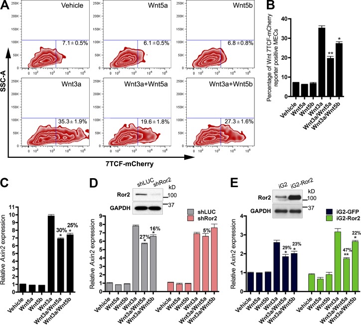
Wnt/β-catenin–dependent and –independent pathways are integrated in the mammary epithelium. (A) FACS profiles of primary MECs representing the percentage of 7TCF-mCherry induction after treatment with Wnt3a, Wnt5a, and Wnt5b. Shown are representative FACS plots of three experiments, where treatments were performed in triplicate. (B) Bar graph representing the percentage of Wnt/β-catenin–dependent induction in A by Wnt3a and the degree of inhibition by either Wnt5a or Wnt5b. (C) qRT-PCR analysis of Axin2 induction after Wnt stimulation. (D) qRT-PCR analysis of Axin2 after Wnt stimulation in shLUC and shRor2 primary MECs. Depletion of Ror2 expression by shRNA-mediated silencing (illustrated by the Western blot inset) reduced both Wnt5a and Wnt5b antagonism of Axin2 by Wnt3a. (E) qRT-PCR analysis of Axin2 in response to Wnt treatments after Ror2 overexpression. Ror2 expression above steady-state levels (illustrated by the Western blot inset) enhanced Wnt/β-catenin pathway repression by Wnt5a (iG2 29% and iG2-Ror2 47% inhibition), but no difference was observed for Wnt5b (iG2 23% and iG2-Ror2 22% inhibition). Plotted values represent means ± SD (error bars). *, P < 0.05; **, P < 0.01.

