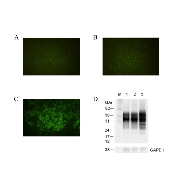
Figure 1. Immunofluorescence and Immunoblotting detection of bovine cellular prion protein (PrPC) in transduced/non-transduced Madin-Darby bovine kidney (MDBK; ATCC CCL-22) with infectious recombinant lentivirus.

Detection of cell surface PrPC in non-transduced MDBK (A), transduced MDBK with only pLEX vector (B) and MDBK C1–2F with bovine PRNP inserted into pLEX vector (C) were assayed by immunofluorescence using 4% paraformaldehyde fixation and anti-PrP mAb 6H4. (D) This photo shows an immunoblot of non-transduced MDBK (lane 1) and transduced MDBKs with infectious recombinant virus which had pLEX vector without and with bovine PRNP (lane 2, 3). Housekeeping protein, GAPDH (glyceraldehydes-3-phosphate dehydrogenase) was used as a control of comparative protein concentration. The positions of molecular marker proteins (M) are presented in kilodaltons.
