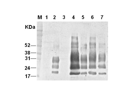
Figure 4. Immunoblotting detection of the persistent PrPBSE-infected cell according to serial passages.

The lysates of brain homogenates (lane 1; BSE-noninfected bovine brain, lane 2; BSE-infected bovine brain-original BSE source) and MDBK cells expressing normal bovine prion protein by using infectious recombinant lentivirus (lane 3: transduced MDBK–MBDK C1–2F–passage(p) 7; lane 4: BSE-infected transduced MDBK (M2B)–p17; lane 5: M2B-p48; lane 6: M2B-p70 and lane 7: M2B-p83) were treated with proteinase K (PK) for detection of PrPBSE in transduced MDBK which was sequentially passaged after inoculating BSE-infected bovine brain homogenate. Molecular mass marker (M) in kilodaltons (kDa) is shown on the left. The result shown is representative of multiple independent experiments.
