
Fig 2. Cholinergic Neurons of the Laterodorsal Tegmental Nucleus in the Ventrolateral Periaqueductal Gray.
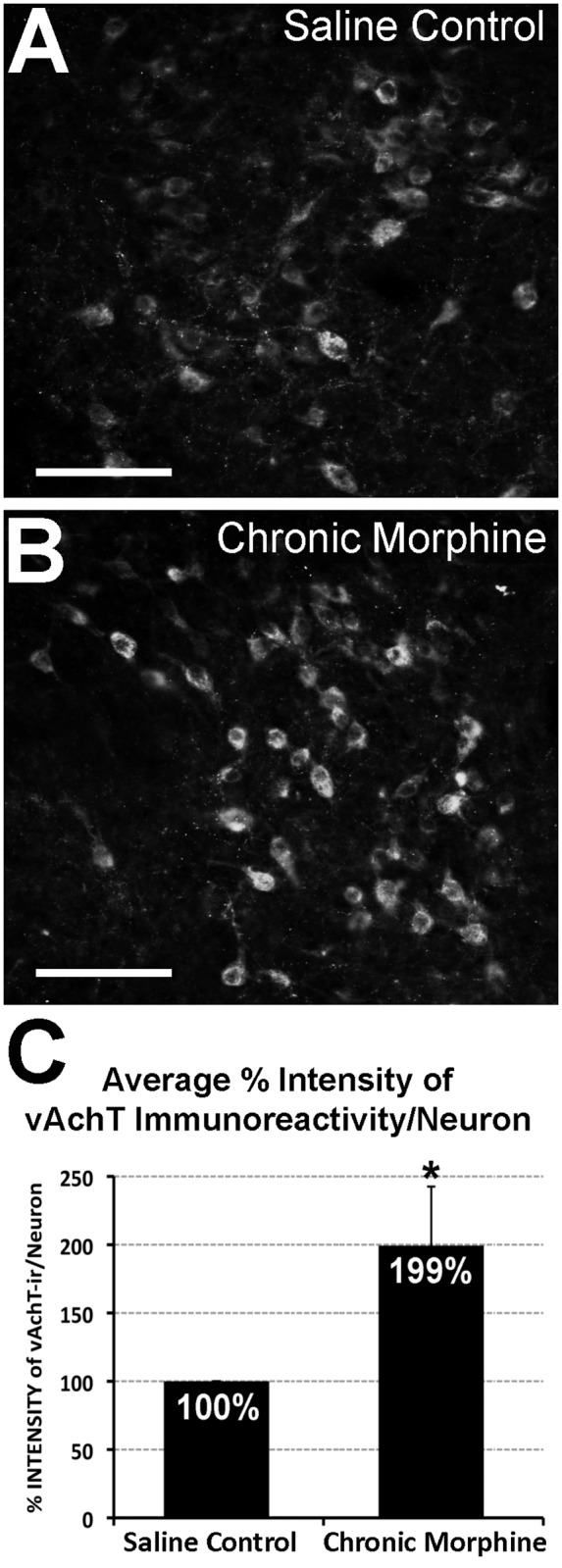
Photomicrographs illustrate vesicular acetylcholine transporter (vAChT) immunofluorescence in the laterodorsal tegmental nucleus. Cholinergic neurons are labeled more intensely following chronic morphine treatment (B) in comparison to saline control group (A). Graph in Panel C illustrates average percentage (%) intensity of vAChT immunoreactivity per individual cholinergic neuron in the laterodorsal tegmental nucleus (± SD; n = 6/group). There is a statistically significant increase (*) in intensity of vAChT immulabeling/neuron following chronic morphine administration when compared to control (99% ± 43.31; t(10) = -5.61, two-tailed p<0.001). Scale bar = 100 μm.