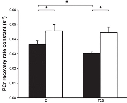
Fig. 2.

Exercise restores mitochondrial function in type 2 diabetic (T2D) subjects. In vivo mitochondrial function was measured in vastus lateralis muscle and expressed as the rate constant (in s−1) before (solid bars) and after training (open bars). A high rate constant reflects high in vivo mitochondrial function. Pre- and posttraining leg extension exercise was performed at 0.5 Hz to an acoustic cue on a magnetic resonance-compatible ergometer and a weight corresponding to 60% of the predetermined maximum. Postexercise phosphocreatine (PCr) resynthesis is driven almost purely oxidatively, and the resynthesis rate reflects in vivo mitochondrial function. Data are expressed as means ± SE. #Data for T2D subjects were significantly different from those of the control (C) group. *Posttraining was significantly different from pretraining. [Adapted from Ref. 52.]
