
Fig. 3.
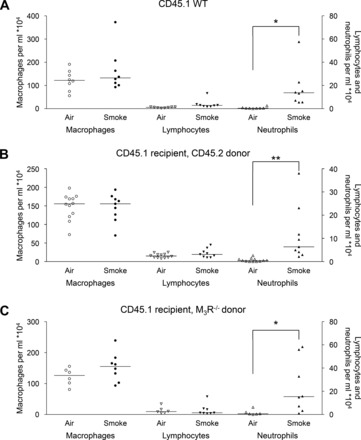
Inflammatory cells in the lavage fluid of CD45.1 animals. Mice were treated as described in Fig. 1. Sixteen hours after the last smoke exposure a bronchoalveolar lavage was performed, and macrophages, lymphocytes, and neutrophils were determined in the lavage fluid of nonirradiated wild-type (WT) animals (A), wild-type animals receiving wild-type bone marrow cells (B), and wild-type animals receiving M3 receptor-deficient (M3R−/−) bone marrow cells (C). *P < 0.05 and **P < 0.01; n = 6–12 animals.