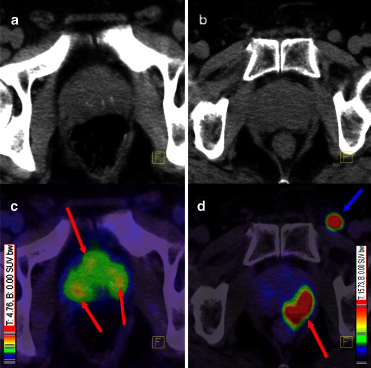
Fig. 2.
A patient with multifocal PCa (a, c) and another patient (b, d) with unifocal PCa with a rarely seen inguinal lymph node metastasis. Red arrows point to PCa within the prostate gland and blue arrow points to an inguinal lymph node metastasis. Both patients had GSC 7, although the tumours present with different contrast. Colour scales were automatically produced by the PET/CT machine. a Low-dose CT of the patient with a multifocal PCa, c corresponding fusion of PET and low-dose CT 1 h p.i., b low-dose CT of the patients with the unifocal PCa, d corresponding fusion of PET and low-dose CT 1 h p.i.

