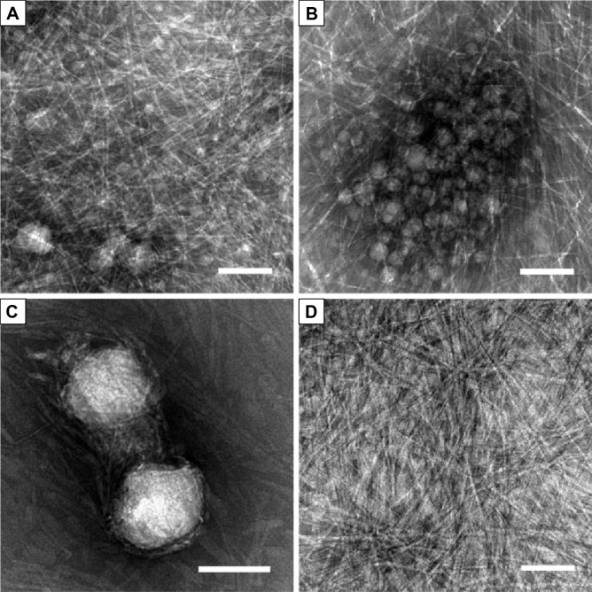
Figure 3.
Transmission electron microscopic images of nanostructures in the suspension and supernatant.
Notes: (A–C) Nanostructures in the suspension. (A, B) were from the same sample, showing A6K nanofibers as well as nanoparticles with a diameter of tens of nanometers. (C) Larger pyrene particles with a diameter of about 100 nm were observed in another transmission electron microscopy sample. (D) Only A6K nanofibers were present in the supernatant. Scale bar, 100 nm.

