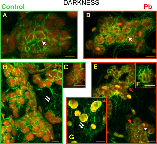
Fig 7. Visualization of actin filaments Lemna trisulca mesophyll cells.
With the help of Alexa Fluor 488—phalloidin (green fluorescence) in control plants (A-C) and plants treated with lead (D-G) in darkness. Chloroplasts are visible thanks to the red autofluorescence of chlorophyll. The network of microfilament bundles varying in thickness, which were not directly connected with chloroplasts, is marked with double arrows. Microfilament bundles twisting around plastids (single white arrows), with their branches, are also marked (C and F). In cells treated with lead, disturbances in microfilament pattern were recorded: absence of long bundles twisting around plastids (asterisk), fragmentation of bundles twisting around chloroplasts (red arrow), and thick, local accumulations of actin (red arrowhead). Scale bar = 5 μm

